- Anatomy
- Conditions
- Procedures
- Others
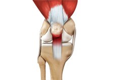
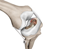
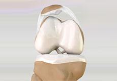
Knee Anatomy
The knee is a complex joint made up of different structures - bones, tendons, ligaments, and muscles. They all work together to maintain the knee’s normal function and provide stability to the knee during movement.
Having a well-functioning healthy knee is essential for our mobility and ability to participate in various activities. Understanding the anatomy of the knee enhances your ability to discuss and choose the right treatment procedure for knee problems with your doctor.
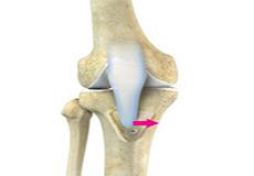
Bones of the Knee
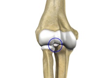
The knee is a hinge joint made up of two bones, the thighbone (femur) and shinbone (tibia). There are two round knobs at the end of the femur called femoral condyles that articulate with the flat surface of the tibia called the tibial plateau. The tibial plateau on the inside of the leg is called the medial tibial plateau and on the outside of the leg, the lateral tibial plateau.
The two femoral condyles form a groove on the front (anterior) side of the knee called the patellofemoral groove. A small bone called the patella sits in this groove and forms the kneecap. It acts as a shield and protects the knee joint from direct trauma.
A fourth bone called the fibula is the other bone of the lower leg. This forms a small joint with the tibia. This joint has very little movement and is not considered a part of the main joint of the knee.
Articular Cartilage and Menisci of the Knee
Movement of the bones causes friction between the articulating surfaces. To reduce this friction, all articulating surfaces involved in the movement are covered with a white, shiny, slippery layer called articular cartilage. The articulating surface of the femoral condyles, tibial plateaus and the back of the patella are covered with this cartilage. The cartilage provides a smooth surface that facilitates easy movement.
To further reduce friction between the articulating surfaces of the bones, the knee joint is lined by a synovial membrane that produces a thick clear fluid called synovial fluid. This fluid lubricates and nourishes the cartilage and bones inside the joint capsule.
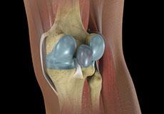
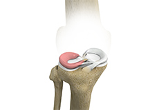
Within the knee joint, between the femur and tibia, are two C-shaped cartilaginous structures called menisci. Menisci function to provide stability to the knee by spreading the weight of the upper body across the whole surface of the tibial plateau. The menisci help in load-bearing i.e. it prevents the weight from concentrating onto a small area, which could damage the articular cartilage. The menisci also act as a cushion between the femur and tibia by absorbing the shock produced by activities such as walking, running and jumping.
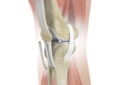
Ligaments of the Knee
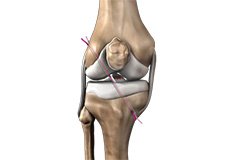
Ligaments are tough bands of tissue that connect one bone to another bone. The ligaments of the knee stabilize the knee joint. There are two important groups of ligaments that hold the bones of the knee joint together, collateral and cruciate ligaments.
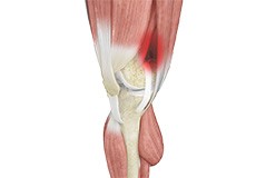
Collateral ligaments are present on either side of the knee. They prevent the knee from moving too far during side to side motion. The collateral ligament on the inside is called the medial collateral ligament (MCL) and the collateral ligament on the outside is called the lateral collateral ligament (LCL).
Cruciate ligaments, present inside the knee joint, control the back-and-forth motion of the knee. The cruciate ligament in the front of the knee is called anterior cruciate ligament (ACL) and the cruciate ligament in the back of the knee is called posterior cruciate ligament (PCL).
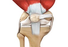
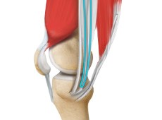
Muscles of the Knee
There are two major muscles in the knee - the quadriceps and the hamstrings, which enable movement of the knee joint. The quadriceps muscles are located in front of the thigh. When the quadriceps muscles contract, the knee straightens. The hamstrings are located at the back of the thigh. When the hamstring muscles contract, the knee bends.
Tendons of the Knee
A tendon is a tissue that attaches a muscle to a bone. The quadriceps muscles of the knee meet just above the patella and attach to it through a tendon called the quadriceps tendon. The patella further attaches to the tibia through a tendon called the patella tendon. The quadriceps muscle, quadriceps tendon, and patellar tendon all work together to straighten the knee. Similarly, the hamstring muscles at the back of the leg are attached to the knee joint with the hamstring tendon.
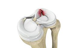
Knee Fracture
A fracture is a condition in which there is a break in the continuity of the bone. In younger individuals, these fractures are caused by high energy injuries, as from a motor vehicle accident. In older people, the most common cause is a weak and fragile bone.
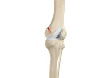
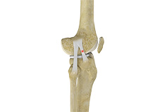
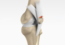
ACL Tears
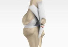
The anterior cruciate ligament (ACL) is one of the major ligaments of the knee. It is located in the middle of the knee and runs from the femur (thighbone) to the tibia (shinbone). The ACL prevents the tibia from sliding out in front of the femur. Together with the posterior cruciate ligament (PCL), it provides rotational stability to the knee.
MCL Sprains
Your MCL may get sprained or injured while twisting, bending or quickly changing direction. MCL sprains occur due to a sudden impact from the outside of your knee, most commonly while playing sports such as rugby and football. Rarely, the MCL can get injured when the knee gets twisted or following a quick change in direction.
Jumper's Knee
Jumper’s knee, also known as patellar tendinitis, is inflammation of the patellar tendon that connects your kneecap (patella) to your shinbone. This tendon helps in the extension of the lower leg.
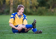
Knee Injury
A knee injury can affect any of the ligaments, tendons or fluid-filled sacs (bursae) that surround your knee joint as well as the bones, cartilage and ligaments that form the joint itself. Some of the more common knee injuries include: ACL injury.
Knee Sprain
Knee sprain is a common injury that occurs from overstretching of the ligaments that support the knee joint. A knee sprain occurs when the knee ligaments are twisted or turned beyond its normal range, causing the ligaments to tear.
Ligament Injuries
Ligament injuries are normally related to trauma that overstresses the ligament beyond its load capacity.
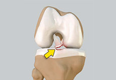
Meniscal Injuries
Injuries to the crescent-shaped cartilage pads between the two joints formed by the femur (the thigh bone) and the tibia (the shin bone). The meniscus acts as a smooth surface for the joint to move on. The two menisci are easily injured by the force of rotating the knee while bearing weight.
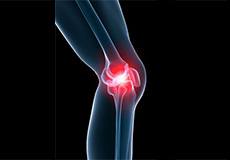
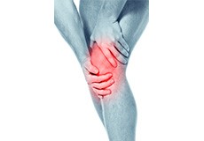
Knee Pain
Knee pain is a common condition affecting individuals of various age groups. It not only affects movement but also impacts your quality of life. An injury or disease of the knee joint or any structure surrounding the knee can result in knee pain. A precise diagnosis of the underlying cause is important to develop an appropriate treatment plan.
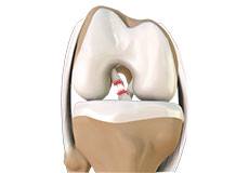
Meniscal Tears
A meniscal tear is a common knee injury in athletes, especially those involved in contact sports. A sudden bend or twist in your knee causes the meniscus to tear. Elderly people are more prone to degenerative meniscal tears as the cartilage wears out and weakens with age.
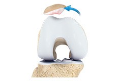
Fractures of the Patella
The patella or kneecap is a small bone present in the front of your knee where the thigh bone meets the shinbone. It provides protection to your knee and attachment to muscles in the front of the thigh. An injury to the knee can result in a break or fracture of the patella.
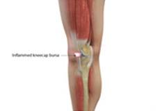
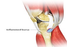
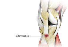
Kneecap Bursitis
Bursitis refers to the inflammation and swelling of the bursa. Inflammation of the bursa in front of the kneecap (patella) is known as kneecap bursitis or prepatellar bursitis.
Runner's Knee
Patellofemoral pain syndrome also called runner’s knee refers to pain under and around your kneecap. Patellofemoral pain is associated with a number of medical conditions such as anterior knee pain syndrome, patellofemoral malalignment, and chondromalacia patella.
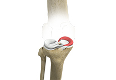
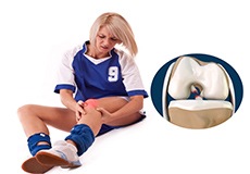
Patellar Instability
Any damage to the supporting ligaments may cause the patella to slip out of the groove either partially (subluxation) or completely (dislocation). This misalignment can damage the underlying soft structures such as muscles and ligaments that hold the kneecap in place.
Patella Fracture
The kneecap or patella forms a part of the knee joint. It is present at the front of the knee, protecting the knee and providing attachment to various muscle groups of the thigh and leg.
Knee Arthritis
The joint surface is covered by a smooth articular surface that allows pain-free movement in the joint. Arthritis is a general term covering numerous conditions where the joint surface or cartilage wears out. This surface can wear out for several reasons; often the definite cause is not known. Arthritis often affects the knee joint. When the articular cartilage wears out, the bone ends rub on one another and cause pain.
Lateral Patellar Compression Syndrome
Lateral patellar compression syndrome refers to pain under and around your kneecap. It is a common complaint among runners, jumpers and other athletes such as skiers, cyclists, and soccer players.
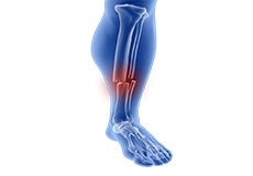
Fractures of the Tibia
The lower leg is made up of two long bones called the tibia and fibula that extend between the knee and ankle. The tibia or shinbone is the larger of the two bones. It bears most of the body’s weight and helps form the ankle joint and knee joint.
Chondromalacia Patella
Chondromalacia patella is a common condition characterized by softening, weakening and damage of the cartilage. The condition is most often seen in young athletes and older adults who have arthritis of the knee. It especially occurs in women.
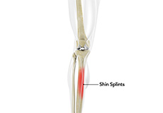
Shin Splints
Shin splints are pain and inflammation of the tendons, muscles and bone tissue along the tibia or shinbone (lower leg). It occurs because of vigorous physical activities such as exercise or sports. The condition is also referred to as medial tibial stress syndrome (MTSS).
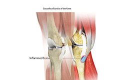
Goosefoot Bursitis of the Knee
A bursa is a small fluid-filled sac found between soft tissues and bones. It lubricates and acts as a cushion, decreasing the friction between bones when they move. Bursitis refers to the inflammation and swelling of the bursa. Goosefoot bursitis or pes anserine bursitis is the inflammation of the bursa present between the tendons of the hamstring muscle and the tibia (shinbone) on the inner side of the knee.
Baker's Cyst
The knee consists of a fluid called synovial fluid, which reduces the friction between the bones of the knee joint while you move your leg. Sometimes this fluid is produced in excess, resulting in its accumulation in the back of your knee. A Baker’s cyst or popliteal cyst is a fluid-filled swelling that develops into a lump behind the knee. This causes stiffness, tightness, and pain behind your knee.
Patellar Tendon Rupture
The patellar tendon works together with the quadriceps muscle and the quadriceps tendon to allow your knee to straighten out. Patella tendon rupture is the rupture of the tendon that connects the patella (kneecap) to the top portion of the tibia (shinbone).
Iliotibial Band Syndrome
Iliotibial band syndrome is an overuse injury resulting from the inflammation of the iliotibial band. It occurs when the iliotibial band and the lower outside portion of the thighbone at the knee joint rub against each other.
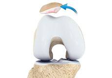
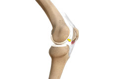
Patellar Dislocation/Patellofemoral Dislocation
Patellar dislocation occurs when the patella moves out of the patellofemoral groove, (trochlea) onto the bony head of the femur. If the kneecap partially comes out of the groove, it is called subluxation; if the kneecap completely comes out, it is called dislocation (luxation).

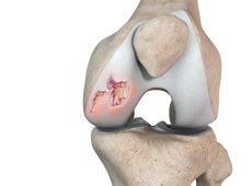
Osteochondritis Dissecans of the Knee
Osteochondritis dissecans is a joint condition in which a piece of cartilage, along with a thin layer of the bone separates from the end of the bone because of inadequate blood supply. The separated fragments are sometimes called “joint mice”.
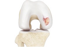
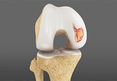
Chondral or Articular Cartilage Defects
The articular or hyaline cartilage is the tissue lining the surface of the two bones in the knee joint. Cartilage helps the bones move smoothly against each other and can withstand the weight of your body during activities such as running and jumping.
Medial Meniscus Syndrome
>Of the menisci within the knee, it is the medial that is more easily injured. Differences in the anatomical attachments of the medial meniscus compared to the lateral, mean that the medial meniscus becomes distorted during combined flexion and rotation movements in a manner not experienced on the lateral side.
Patellar Tendinitis
Patellar tendinitis, also known as "jumper's knee", is an inflammation of the patellar tendon that connects your kneecap (patella) to your shinbone. This tendon helps in extension of the lower leg.
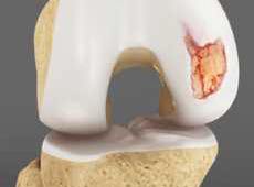
Articular Cartilage Injury
Articular or hyaline cartilage is the tissue lining the surface of the two bones in the knee joint. Cartilage helps the bones move smoothly against each other and can withstand the weight of the body during activities such as running and jumping.
Recurrent Patella Dislocation
The patella (kneecap) is a small bone that shields your knee joint. It is present in front of your knee, on a groove called the trochlear groove that sits at the junction of the femur (thighbone) and tibia (shinbone).
Quadriceps Tendon Rupture
The quadriceps can rupture after a fall, direct blow to the leg and when you land on your leg awkwardly from a jump. Quadriceps tendon rupture most commonly occurs in middle-aged people who participate in sports that involve jumping and running.
Lateral Meniscus Syndrome
Lateral meniscus syndrome is characterized by an injury caused by the tearing of the cartilage tissue or a rare case of a congenital abnormality called a discoid meniscus, which results in knee pain.
Osteonecrosis of the Knee
Osteonecrosis is a condition in which the death of a section of bone occurs because of lack of blood supply to it. It is one of the most common causes of knee pain in older women.

Osteochondral Defect of the Knee
An osteochondral defect, also commonly known as osteochondritis dissecans, of the knee refers to a damage or injury to the smooth articular cartilage surrounding the knee joint and the bone underneath the cartilage.
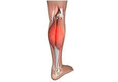
Medial Gastrocnemius Strain
A medial gastrocnemius strain (MGS), also sometimes called “tennis leg”, is an injury to the calf muscle in the back of the leg. It occurs when the calf muscle is stretched too far resulting in a partial or total tear or rupture within the muscle.
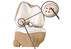
Loose Bodies in the Knee
Loose bodies are fragments of detached cartilage or bone inside the knee joint. These fragments may be free floating (unstable) or may be trapped (stable) within the joint. Depending on the severity, you may have one or more loose bodies in your knee joint.
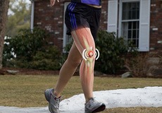
Knee Sports Injuries
Trauma is any injury caused during physical activity, motor vehicle accidents, electric shock, or other activities. Sports trauma or sports injuries refer to injuries caused while playing indoor or outdoor sports and exercising. Sports trauma can result from accidents, inadequate training, improper use of protective devices, or insufficient stretching or warm-up exercises.
Pediatric Tibial Tubercle Fractures
Tibial tubercle fractures are quite rare occurrences that typically affect physically active adolescents between the age of 14 and 17. It is caused by violent tensile forces exerted over the tibial tuberosity (a bulge in the tibial bone) during activities involving sudden contraction of the knee extensors (springing and jumping).
Women and ACL Injuries
The anterior cruciate ligament is one of the four major ligaments of the knee that connects the femur (thigh bone) to the tibia (shin bone) and helps stabilize the knee joint. Anterior cruciate ligament (ACL) injury is one of the common injuries of the knee.
Adolescent Knee Problems
Osgood-Schlatter disease is a common knee problem seen in growing adolescents.Osgood-Schlatter disease refers to a condition in older children and teenagers caused by excessive stress to the patellar tendon (located below the kneecap). Participants in sports such as soccer, gymnastics, basketball, and distance running are at higher risk for this disease.
Anterior Knee Pain
Anterior knee pain is characterized by chronic pain over the front and center of the knee joint. It is common in athletes, active adolescents (especially girls) and overweight individuals.
Osgood-Schlatter Disease
Osgood-Schlatter disease refers to an overuse injury that occurs in the knee of growing children and adolescents. This is caused by inflammation of the tendon located below the kneecap (patellar tendon). Children and adolescents who participate in sports such as soccer, gymnastics, basketball, and distance running are at a higher risk of this disease.
Tibial Eminence Spine Avulsion Fracture
Tibial eminence spine avulsion fracture is the avulsion (tearing away) of the tibial eminence.
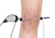
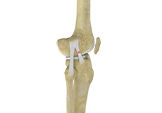
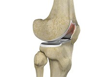
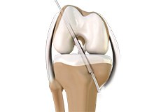
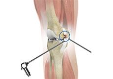
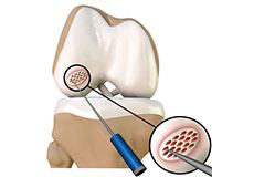
Knee Arthroscopy
Knee arthroscopy is a surgical technique that can diagnose and treat problems in the knee joint. During the procedure, your surgeon will make a very small incision and insert a tiny camera — called an arthroscope — into your knee. This allows them to view the inside of the joint on a screen.
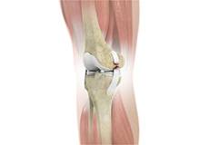
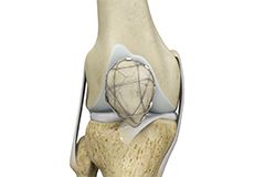
ACL Reconstruction
The anterior cruciate ligament (ACL) is one of the major stabilizing ligaments in the knee. It is a strong rope-like structure located in the center of the knee, running from the femur to the tibia.
Meniscus Repair
A meniscus tear is the commonest knee injury in athletes, especially those involved in contact sports. A sudden bend or twist in your knee can cause the meniscus to tear. This is a traumatic meniscal tear. The elderly is more prone to degenerative meniscal tears as the cartilage wears out and weakens with age.
Meniscal Surgery
Two wedge-shaped cartilage pieces are present between the thighbone and shinbone. These are called menisci. They stabilize the knee joint and act as shock absorbers.
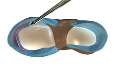
Cartilage Replacement
Cartilage replacement is a surgical procedure performed to replace the worn-out cartilage with new cartilage.
Multiligament Reconstruction
Multiligament knee reconstruction is a surgical procedure to repair or replace two or more damaged ligaments of the knee joint. The surgery can be performed using minimally invasive techniques.
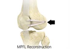
Medial Patellofemoral Ligament Reconstruction
The medial patellofemoral ligament is a band of tissue that extends from the femoral medial epicondyle to the superior aspect of the patella. It is a major ligament that stabilizes the patella and helps in preventing patellar subluxation (partial dislocation) or dislocation.
Arthroscopic Reconstruction of the Knee for Ligament Injuries
Arthroscopic reconstruction of the knee ligament is a minimally invasive surgery performed through a few tiny incisions. An arthroscope is inserted into the knee joint through one of the small incisions to provide clear images of the surgical area (inside the knee) to your surgeon on a television monitor.
Patellar Tendon Repair
Patella tendon rupture is the rupture of the tendon that connects the patella (kneecap) to the top portion of the tibia (shinbone). The patellar tendon works together with the quadriceps muscle and the quadriceps tendon to allow your knee to straighten out.
Quadriceps Tendon Repair
Quadriceps tendon is a thick tissue located at the top of the kneecap. The quadriceps tendon works together with the quadriceps muscles to allow us to straighten our leg. The quadriceps muscles are the muscles located in front of the thigh.
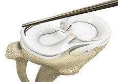
Partial Arthroscopic Meniscectomy
Partial arthroscopic meniscectomy is a procedure to remove the damaged part of a meniscus in the knee joint with the help an arthroscope. The meniscus is a C-shaped disc of cartilage between your thighbone and shinbone. There are 2 menisci in each knee. They act as shock absorbers and provide stability to the joint.
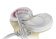
Saucerization
Saucerization is a surgical procedure performed to treat a discoid (disc-shaped) meniscus in the knee joint which is more prone to injury. The normal meniscus is crescent-shaped cartilage cushioning the ends of the femur (thighbone) and tibia (shinbone) in the knee.
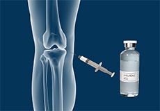
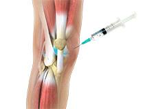
Intraarticular Knee Injection
An intra-articular knee injection is a very effective form of treatment where medicine is delivered directly into the knee joint with the primary objective of relieving pain from conditions such as arthritis.
Combined Hyaluronic Therapy for the Knee
Combined hyaluronic therapy is the process of injecting hyaluronic acid (HA) along with platelet-rich plasma (PRP) into your knee to treat osteoarthritis.
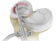
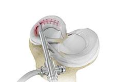
Matrix Induced Autologous Chondrocyte Implantation (MACI)
Matrix-Induced autologous chondrocyte implantation is an innovative, FDA-approved cartilage restoration procedure that uses your own cells to repair cartilage defects in your knee. It can alleviate knee pain, help you regain function and may even delay or prevent arthritis.
Arthroscopic Debridement
Arthroscopic debridement or a clean-up is a surgical procedure performed using an arthroscope. In this procedure, the cartilage or the bone that is damaged is removed using surgical instruments and the edges of the articular cartilage that are rough will be smoothened.
Failed Anterior Cruciate Ligament (ACL) Reconstruction
The knee joint is stabilized by four strong ligaments. The anterior cruciate ligament (ACL) passes diagonally in the middle of the knee, ensuring that the thigh and shin bone do not slide out of alignment during movement. ACL injury is one of the most common sports injuries of the knee joint and is generally repaired with ACL reconstruction surgery, where the torn ligament is replaced with a graft tendon.
Failed Meniscus Repair
Meniscal repair may be performed either by open surgery under direct vision or minimally invasively using an arthroscope, which is a thin tube fitted with a camera that can be inserted into the knee through a very small incision to locate and repair the damaged meniscus.
Meniscectomy
Meniscectomy is a surgical procedure indicated in individuals with torn meniscus where the conservative treatments are a failure to relieve the pain and other symptoms. Meniscectomy is recommended based on the ability of meniscus to heal, patient’s age, health status, and activity level.
Mosaicplasty
Mosaicplasty is a surgical technique to repair the defect by transplanting healthy bone and cartilage from non-weight bearing areas of the knee. It is indicated to treat small cartilage defects of less than 2 cm in young active adults less than 45 years of age.
Prior Meniscectomy
The menisci are two C-shaped cartilages that act as shock absorbers between the thigh and shin bones that articulate at the knee joint. They provide stability and lubrication to the joint as well as nutrition for the articular cartilage. Tears in the meniscus may occur as a result of acute injury or chronic degeneration with age.
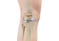
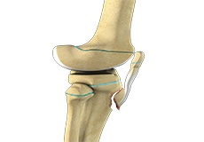
Tibial Eminence Fracture
The tibia or shin bone is a major bone of the leg which connects the knee to the ankle. A fracture or break in the upper part of the tibia is known as a proximal tibial fracture and commonly occurs just below the knee joint.
ORIF of the Knee Fracture
ORIF refers to open reduction and internal fixation. It is a surgical procedure employed for the treatment of a fracture not amenable to non-surgical conservative treatment.
Chondroplasty
Chondroplasty is a surgical procedure to repair and reshape damaged cartilage in a joint. The procedure involves smoothing degenerative cartilage and trimming any unstable flaps of cartilage.
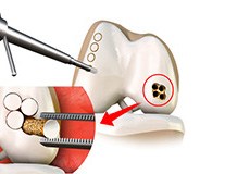
Bone-Patellar Tendon-Bone (BPTB) Autograft
ACL reconstruction with BPTP autograft is a surgical procedure that replaces the injured ACL with an autograft containing patellar tendon and bony attachments. The new ACL is harvested from the patellar tendon that connects the bottom of the kneecap (patella) to the top of the shinbone (tibia).
Bone-Patellar Tendon-Bone (BPTB) Allograft
ACL reconstruction with BPTP autograft is a surgical procedure that replaces the injured ACL with an autograft containing patellar tendon and bony attachments. The new ACL is harvested from the patellar tendon that connects the bottom of the kneecap (patella) to the top of the shinbone (tibia).
Hamstring Autograft
ACL reconstruction with hamstring autograft method is a surgical procedure to replace the torn ACL with part of the hamstring tendon taken from your leg. The goal of ACL reconstruction surgery is to tighten your knee and restore its stability.

Pharmacological Interventions for Knee Injuries
Pharmacological interventions include medicinal preparations such as pain-relieving capsules or injections.
Hamstring Allograft
ACL reconstruction hamstring allograft method is a surgical procedure to replace the torn ACL with hamstring allograft. The goal of ACL reconstruction surgery is to tighten your knee and restore its stability.
Viscosupplementation
Viscosupplementation refers to the injection of a hyaluronan preparation into the joint. Hyaluronan is a natural substance present in the joint fluid that assists in lubrication. It allows the smooth movement of the cartilage-covered articulating surfaces of the joint.

Physical Therapy for Knee
Physical therapy is an exercise program that helps you to improve movement, relieve pain, encourage blood flow for faster healing, and restore your physical function and fitness level. It can be prescribed as an individual treatment program or combined with other treatments.
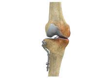
High Tibial Osteotomy
High tibial osteotomy is a surgical procedure performed to relieve pressure on the damaged site of an arthritic knee joint. It is usually performed in arthritic conditions affecting only one side of your knee and the aim is to take pressure off the damaged area and shift it to the other side of your knee with healthy cartilage. During the surgery, your surgeon will remove or add a wedge of bone either below or above the knee joint depending on the site of arthritic damage.
Tibial Tubercle Osteotomy
Tibial tubercle osteotomy is a surgical procedure that is performed along with other procedures to treat patellar instability, patellofemoral pain, and osteoarthritis. The tibial tubercle transfer technique involves realignment of the tibial tubercle (a bump in the front of the shinbone) such that the kneecap (patella) traverses in the center of the femoral groove.
ACL Reconstruction of Patellar Tendon
The anterior cruciate ligament is one of the four major ligaments of the knee that connects the femur (thighbone) to the tibia (shinbone) and helps stabilize the knee joint. The anterior cruciate ligament prevents excessive forward movement of the lower leg bone (tibia) in relation to the thighbone (femur) as well as limits rotational movements of the knee.
ACL Reconstruction Procedure of Hamstring Tendon
The anterior cruciate ligament (ACL) is one of the major stabilizing ligaments in the knee. It is a strong rope-like structure located in the center of the knee running from the femur to the tibia. When this ligament tears, it does not heal on its own and often leads to the feeling of instability in the knee.
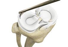
Partial Meniscectomy
Partial meniscectomy is a surgical procedure to remove the torn portion of the meniscus from the knee joint.
Nonoperative Treatments for ACL Injuries
Nonoperative treatments may be recommended if your child has a minor ACL injury, is still growing and does not wish to immediately return to sports.
Non-Surgical Knee Treatments
The knee is a complex joint which consists of bone, cartilage, ligaments, and tendons that make joint movements easy and at the same time it is more susceptible to various kinds of injuries. Knee problems may arise if any of these structures get injured by overuse or suddenly during sports activities.